Quiz: Tingling and Weakness
A 52-year-old female with a history of breast cancer is sent to the ED by her oncologist for a mild headache that's lasted 10 days. What does the EKG show?

")
(©StudioGrandWeb, AdobeStock)


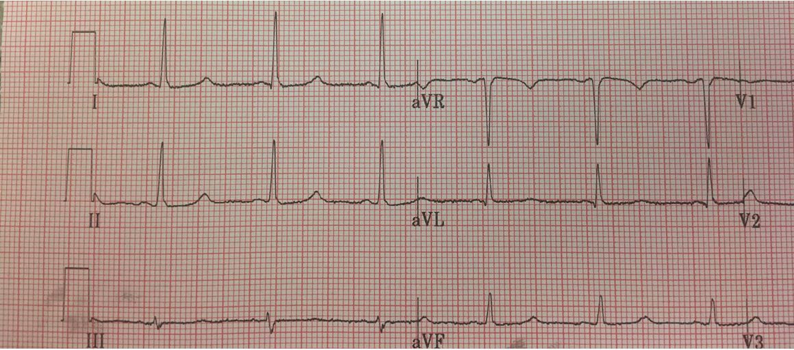
EKG of female patient with a history of breast cancer complaining of headache, weakness, among other complaints. Image courtesy of Dr. Brady Pregerson.


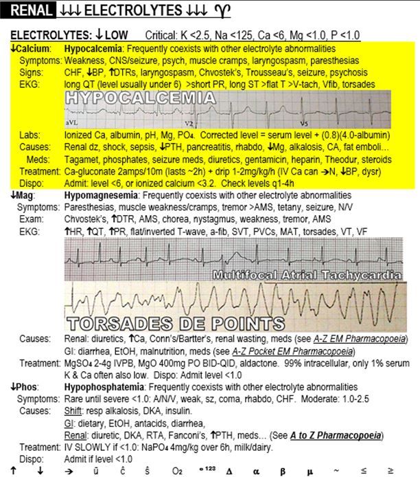
Excerpt on Hypocalcemia from "The Emergency Medicine 1-Minute Consult Pocketbook." Image courtesy of Dr. Brady Pregerson.
History of Present Illness:
A 52-year-old female with a history of breast cancer is sent to the ED by her oncologist to have a brain MRI done to rule out metastases. She has had 2 weeks of progressive generalized weakness, bilateral hand tingling and vomiting that has not responded to stopping her chemo or radiation therapy. She admits to a mild headache on and off for 10 days. She denies any focal weakness, abdominal pain, fever or other complaints.
Vital Signs & Physical Exam:
Vital signs are normal. Physical exam is normal except for minor pronator drift on the right, but the patient complains that she has a bad right shoulder.
Initial Differential Diagnosis:
• Brain mass
• Electrolyte abnormality
• Anxiety
• Medication side effect
Initial Diagnostic Testing:
• The EKG is shown in the image on the right.
What does the case image show?
The EKG is actually almost normal, but the QT interval is a bit long with QTc at 490 milliseconds.
What should you do next?
Order electrolyte levels for potassium, calcium and magnesium. Her calcium came back low at 5.9 (normal 8.5-10.2)
Discussion
Hypocalcemia usually presents with symptoms and signs of CNS irritability such as paresthesias, generalized weakness, muscle cramps, increased deep tendon reflexes and generalized weakness. In severe cases hypotension, seizures and psychosis may even occur.
If an EKG is done, the most common finding is a long QT interval, mostly from prolongation of the ST segment. With more severe derangements of calcium the PR can shorten and the T wave may flatten. Ventricular dysrhythmias may also occur, but are rare. When hypocalcemia is suspected or confirmed, other blood work that may be helpful in determining severing and cause are serum albumin, magnesium and phosphate. The formula for correcting the calcium in low albumin states is shown in the highlighted area of the sample page, as are causes of hypocalcemia, the most common of which are renal disease and hypoparathyroidism.
Treatment for hypocalcemia is with calcium repletion, which can start intravenously with calcium gluconate. Patients with levels below 6 should typically be hospitalized.
Case Conclusion
The patient was admitted. The hypocalcemia was determined to be caused by her cancer. The CT was normal. Once her calcium was repleted, she was sent home with calcium supplementation.
About the Author
Dr. Pregerson is chief editor of http://EMresource.org, an emergency medicine website that includes a free EM ultrasound library, EM cases of the month, EM pocket references and more.
REFERENCE


