An Incidental EKG Finding
A 42-year-old female with no significant past medical history presents to the emergency department for two days of constant non-pleuritic, non-exertional, non-radiating chest pain associated with feeling light-headed. What is the diagnosis?

")
(©Fizkes, AdobeStock.com)


")
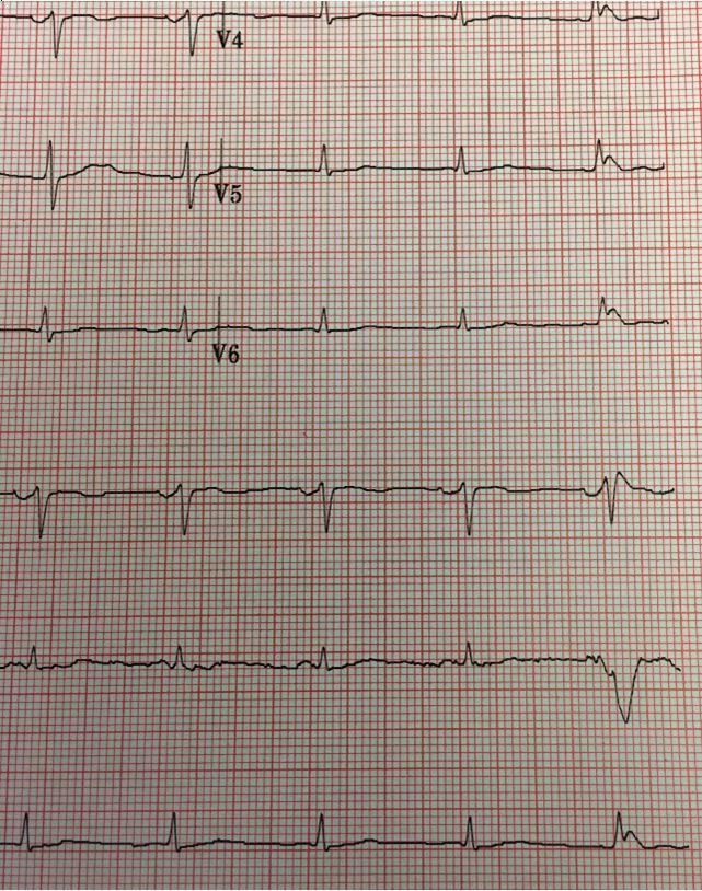
Initial EKG results. (Image courtesy of Dr. Brady Pregerson)


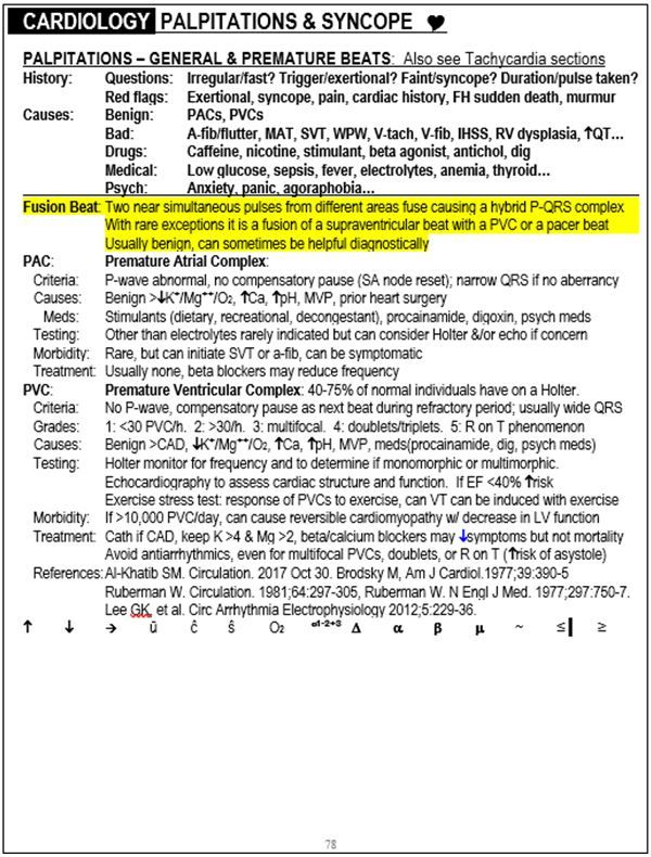
Excerpt on fusion beats from "The Emergency Medicine 1-Minute Consult Pocketbook." Image courtesy of Dr. Brady Pregerson.
History:
A 42-year-old female with no significant past medical history presents to the emergency department for two days of constant non-pleuritic, non-exertional, non-radiating chest pain associated with feeling a bit light-headed. She denies any trouble breathing, cough, palpitations or other complaints
Exam:
Vital signs are normal. Physical exam is normal except for vitiligo. The lungs are clear and heart sounds are normal. Orthostatics are done and are also normal.
Testing:
A screening EKG was done and is shown on the right CBC, metabolic panel and troponin-i are all normal as is the chest X-ray.
See next page for "Computer EKG Results"
Computer EKG Results
1. Sinus rhythm with fusion complexes.
2. Otherwise normal ECG
Do you agree with the computer?
See the next page for the answer.
ANSWERS:
EKG analysis and one-minute consult (Peer-reviewed by Dr. Stephen W. Smith of Dr. Smith’s ECG Blog)
EKG Analysis: The computer read is likely correct. The final beat of the 12 lead appears to be a fusion beat, with a P-wave and a wide QRS different from the intrinsic rhythm. In order to be certain it was a fusion beat and not a PVC, you would need to see a PVC to compare it to.
One-Minute Consult: See highlighted area of the image on the right.
See next page for the discussion.
Discussion
Fusion beats are uncommon and typically benign so are rarely covered in case reports, articles or text books. If you’ve ever wanted to know just a little bit more about them, this is your chance. They occur when an electrical pulse from the atria makes it through the AV node almost simultaneously with an electrical pulse that originates in the ventricle, such as a PVC or ventricular pacer beat. In this way, neither is blocked by a refractory period and both beats fuse giving you the P-wave of a supra-ventricular beat with the wide QRS of a ventricular beat. Sometimes the QRS is of intermediate width. Fusion beats are typically benign but can be helpful diagnostically, such as in cases of ventricular tachycardia.
About the author
Dr. Pregerson is chief editor of http://EMresource.org. an emergency medicine website that includes a free EM ultrasound library, EM cases of the month, EM pocket references and more.


